Rectal hernia

ركتوسل

Introduction

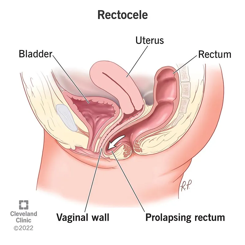
A rectocele occurs when the wall separating the rectum and the vagina (the rectovaginal septum, or the “posterior vaginal wall”) becomes weak and lax, allowing a portion of the rectum to bulge into the vagina. This condition is exclusively a female problem—men do not naturally have a vagina and therefore typically do not develop a rectocele.

Epidemiology (Prevalence and Risk Factors)

  • Rectocele is more common in women, especially in middle age and after menopause.
  • Many women have some degree of pelvic wall weakening, but only a portion of them become symptomatic.
  • Risk increases with the number of vaginal deliveries; long-term constipation, obesity, and chronic coughing also increase the risk.

Etiology (Causes and Mechanisms of the Condition)

Two key processes broadly lead to the development of a rectocele: repeated increases in intra-abdominal pressure and alteration or damage to the supportive tissues.

  1. Repeated intra-abdominal pressure (chronic constipation and straining, chronic coughing, obesity)

  • Each time a person strains, intra-abdominal pressure rises, and consequently the pressure on the pelvic floor and the wall between the rectum and the vagina increases.
  • Recurrent pressure causes continuous stretching of connective tissues: collagen fibers elongate, protein strands are damaged, and their mechanical function gradually diminishes.
  • This stretching is initially reversible, but over time it leads to permanent deformation and thinning of the tissue layers.
  1. Birth-related trauma

  • During difficult or prolonged labor, the rectovaginal septum and pelvic floor muscles undergo significant stretching.
  • There may be tearing of connective tissue fibers or nerve injury. When the tissue is sutured, the repaired area is often less resistant and less elastic, making it prone to future laxity.
  • Use of forceps/vacuum or a large fetus increases the risk of tearing.
  1. Age- and menopause-related tissue changes

  • Decreased estrogen and metabolic changes reduce connective tissue quality and diminish tissue repair capacity.
  1. Connective tissue–related factors

  • Certain rare disorders with inherent collagen weakness (e.g., connective tissue disorders) may increase the risk.
عمل جراحي ركتوپكسي براي پرولاپس ركتوم

Clinical Manifestations

  • A feeling or visible bulge at the back of the vagina when straining.
  • A sensation of incomplete evacuation, with a need to apply manual pressure on the perineum or inside the vagina to aid defecation.
  • Prolonged straining, slow bowel movements, or the need to use a finger for evacuation (splinting).
  • Occasionally, pain or discomfort during sexual intercourse, discharge, or local tenderness.
  • In cases associated with sphincter injury, gas or stool leakage may occur.

Note: The severity of symptoms does not correlate directly with the size of the rectocele; some small rectoceles are highly symptomatic and vice versa.

Diagnosis

History and Examination

  • A detailed history regarding onset, relation to childbirth, need for manual assistance, effects on sexual life, and constipation pattern.
  • Vaginal examination with straining in the supine or standing position; rectal examination to assess sphincter tone and evaluate other lesions.

Fluoroscopic defecography

  • Preparation: Usually partial bowel cleansing the night before; brief explanation and preparation are required.
  • Method: The rectum is filled with a contrast paste resembling stool, and the patient sits on a commode-like chair within the fluoroscopy field. Repeated imaging is performed during rest, straining, and defecation phases.
  • Practical information: Shows the depth and size of the rectocele in millimeters, presence of internal intussusception, multipart or associated rectoceles, and the degree of perineal descent. In practice, a rectocele larger than 20–30 mm (2–3 cm) is often considered clinically significant, but interpretation must be combined with the patient’s symptoms.

Dynamic pelvic MRI

  • Importance: Dynamic MRI provides more comprehensive information about pelvic soft-tissue structures; its major advantage is simultaneous visualization of the rectum, vagina, bladder, levator muscles, and fasciae without ionizing radiation.
  • How it is performed and phases: Typically, the rectum is filled with MRI-compatible gel. The patient is placed in the scanner, and images are obtained in several phases: rest, pelvic floor contraction, straining (Valsalva), and defecation. Some centers have semi-seated setups or special chairs that better simulate real defecation; in all cases, cine MRI (sequential images) displays changes as a moving sequence.
  • Depth of the rectocele (mm) and its type (mucosal vs. full-thickness)
  • Presence of internal intussusception (whether the rectal wall folds inward)
  • Enterocele (herniation of small bowel between rectum and vagina)
  • Perineal descent (mm)
  • Quality and integrity of the levator muscles and presence of scars or muscle tears
  • Organ relationships, such as associated uterine or bladder prolapse
  • Dynamic MRI clarifies whether the problem is a superficial rectocele suitable for vaginal repair, or a multi-compartment/internal issue requiring laparoscopic abdominal treatment.

Manometry and endoanal ultrasound

  • If incontinence or a history of obstetric injury is present, manometry and endoanal ultrasound are essential to evaluate the sphincters and their function. Results influence surgical choice (e.g., adding sphincter repair).

Colonoscopy

  • Before any surgical decision involving the rectum/colon, colonoscopy is required if the patient has not undergone one in recent years, is older than 50, or has alarm symptoms such as unusual bleeding, weight loss, or a family history of cancer, to rule out polyps or tumors. If a recent colonoscopy is available and was normal, repetition is unnecessary during surgical planning.

Treatment

  • If symptoms are mild: constipation management, Kegel exercises, pelvic floor physiotherapy, and toileting behavior training.
  • If symptoms are moderate to severe, or the patient must use “digital splinting,” or suffers from significant sexual pain/discomfort, surgical treatment is considered.
  • Decision-making is best carried out within a multidisciplinary team (colorectal surgeon, gynecologist/perineal specialist, radiologist, physiotherapist).

Non-surgical treatments

  • Diet and stool softeners: gradual fiber intake, polyethylene glycol, or mucosal softeners as needed.
  • Toileting habit training: choosing an appropriate time, avoiding prolonged straining, and using an optimal position (feet on a small stool) to improve the anorectal angle.
  • Kegel exercises and pelvic floor physiotherapy with biofeedback: an 8–12-week program with assessment and guidance from a physiotherapist; if no improvement occurs after an adequate course, treatment should be reassessed.

Kegel exercises (pelvic floor training)

  • Kegels involve voluntary contraction of the pelvic floor muscles (the same muscles you tighten when trying to abruptly stop urination).
  • Correct technique: In a comfortable position (sitting or lying), imagine you are trying to stop urine flow; pull the muscles firmly upward and inward, hold for 5–10 seconds, then relax for 5–10 seconds. Repeat this 8–12 times, three times daily.
  • Tips: Maintain normal breathing during early practice, avoid tightening the buttocks and abdominal muscles, and do not perform the exercise by stopping urine midstream (try that only once to understand the muscle group).
  • When is referral to physiotherapy needed? If you are unsure of the technique, experience pain, or feel no improvement after 6–8 weeks, pelvic floor physiotherapy and biofeedback are beneficial. Biofeedback uses specialized devices to help patients identify the correct muscles and learn appropriate contraction/relaxation patterns.

Surgical Interventions

  1. Posterior Vaginal Repair

  • A small incision is made through the vagina, the fascia and weak tissue are reinforced, and excess mucosa is removed if necessary.
  • Indicated for: Moderate symptomatic rectoceles that involve only the anterior wall, and when MRI/defecography indicates that the problem is limited.
  • Advantages: Direct access, no need for abdominal incision.
  • Limitations: If internal intussusception or multi-organ prolapse is present, the likelihood of insufficient repair and recurrence is higher. In some cases, the use of mesh is controversial, and the risk of mesh exposure and sexual pain should be discussed with the patient.
  1. Stapled Transanal Rectal Resection (STARR)

  • Excess mucosa/submucosa is removed from inside the anal canal, and the area is sutured to reduce protrusion.
  • Indicated for: Patients with primarily evacuation problems due to internal intussusception or excessive mucosal volume.
  • Limitations: Not suitable for full-thickness rectoceles or very large rectoceles; complications such as pain, bleeding, or need for repeat surgery have been reported.
  1. Ventral Rectopexy via Laparoscopy or Robotic Surgery (Minimally Invasive Abdominal Approach)

  • Through an abdominal route, the anterior rectum is lifted with a laparoscopic camera and fixed to a stronger structure with mesh or sutures to correct both prolapse and rectocele.
  • In laparoscopic/robotic techniques, there is better visualization and access to pelvic anatomy.
  • The technique allows for the protection of pelvic nerves, thus reducing the risk of urinary and sexual dysfunction.
  • Simultaneous multi-organ correction (e.g., enteroceles or rectal intussusception) can be performed with a single approach.
  • Less pain and quicker recovery compared to open surgery.
  • Indicated for: Large rectoceles, complete rectal prolapse, cases with multi-organ prolapse, or patients with a history of failed vaginal repair.
  1. Perineal Approach

  • In very elderly or high-risk patients for whom abdominal surgery is not suitable, a small portion is removed through the perineum (in front of the anus), and repair is performed. This is a less invasive approach, but with a higher likelihood of recurrence.

Role of the Colorectal Surgeon

  • Complete evaluation, selection of the appropriate method, performing neuroprotective techniques, and managing complications.
  • Team-based decision-making: Collaboration with a gynecologist (if uterine prolapse or cystocele is present), pelvic floor physiotherapist for pre/post-operative rehabilitation, and radiologist for accurate dynamic MRI reporting is essential.
  • Regarding decisions about childbirth after repair or sexual issues, team consultation and providing the patient with accurate information are necessary.

Frequently Asked Questions about Rectocele

Does Every Rectocele Require Surgery?

No. If the symptoms are mild, non-surgical measures are often sufficient. Surgery is considered when symptoms are present and conservative methods have not been successful.

Because it provides both functional and anatomical information simultaneously: the depth of the rectocele, intussusception, enteroceles, levator muscle status, and perineal descent — all of these are critical for selecting the appropriate surgical method.

Typically, results can be seen within 6–12 weeks with regular practice. If there is no progress after this period, a physiotherapy evaluation is necessary.

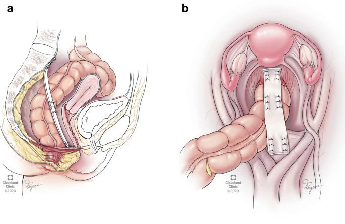
In many cases (especially for large rectoceles or those with multi-organ prolapse), laparoscopy has clear advantages: less pain, quicker recovery, and better nerve preservation. However, the “best” method always depends on the patient’s condition and the surgeon’s experience.

Yes, if the patient has not had a recent colonoscopy, is over 50 years old, or has warning signs, it is necessary to rule out any lesions that may affect the treatment plan.

Summary for Patients

Rectocele refers to the bulging of the front of the rectum into the vagina due to weakness in the supportive wall. Many patients improve with simple treatments like constipation management and pelvic floor exercises. In symptomatic cases, or when the patient needs “manual assistance” for bowel movements, surgical treatment is considered. Dynamic MRI and defecography help in precisely identifying the problem, allowing the best approach (often laparoscopic in complex cases) to be chosen. Team-based decision-making and surgery performed by an experienced center and surgeon yield the best outcomes.