Rectal Cancer

آنوسکوپی

Introduction

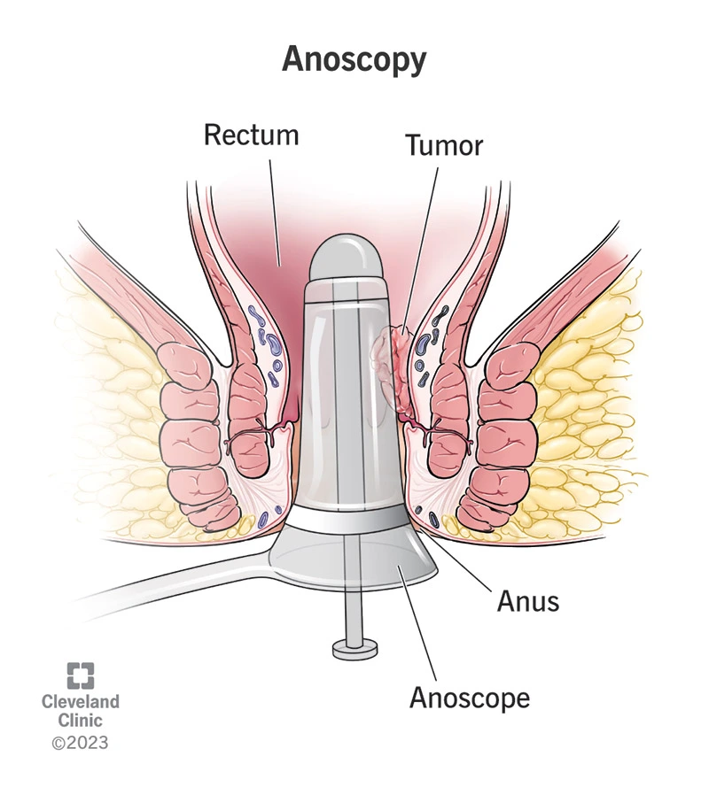
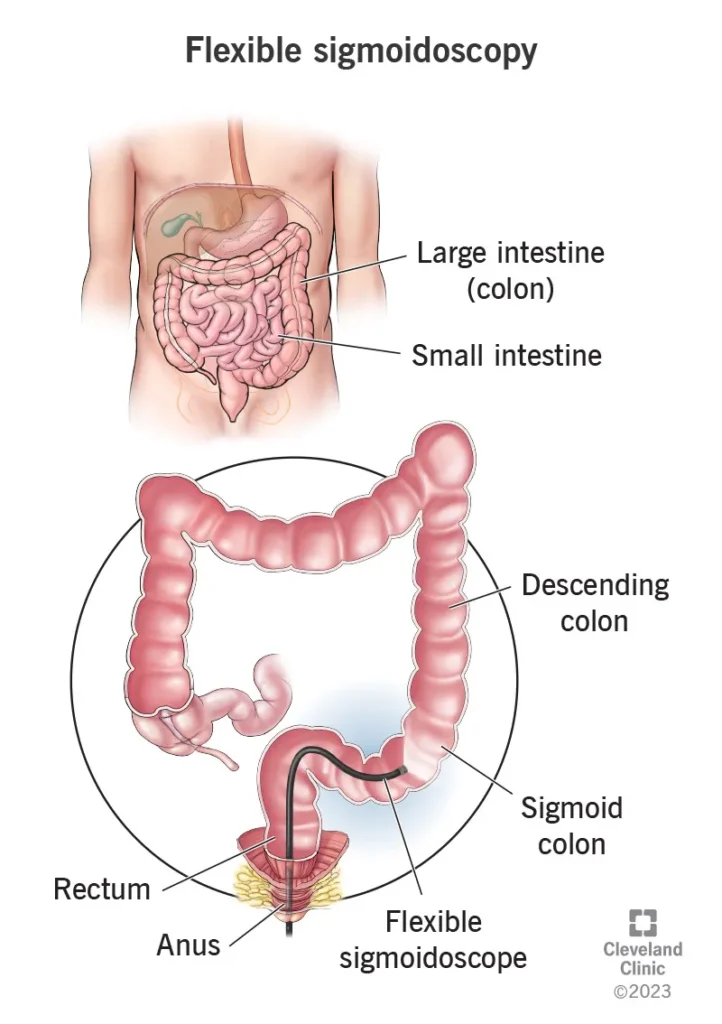
The rectum is the final part of the large intestine, located about 10 to 15 centimeters above the anus. Rectal cancer refers to the uncontrolled growth of malignant cells in this area. Because the rectum is situated within the confined space of the pelvis and lies close to the nerves, bladder, vagina, and reproductive structures, its treatment is technically more complex. Management often requires a combination of radiation therapy, chemotherapy, and surgery, all coordinated by a multidisciplinary medical team.

Epidemiology (Prevalence and Risk Factors)

Rectal cancer accounts for about 30–35% of all colorectal cancers worldwide and is considered one of the major challenges in colorectal surgery. In Western countries, its incidence has been declining due to widespread colonoscopy screening programs. However, in developing countries—including Iran—the incidence continues to rise.

In Iran, the proportion of rectal cancer relative to all colorectal cancers is similar to global statistics, but a notable characteristic is the younger age of onset. A significant number of Iranian patients are under 50 years old at the time of diagnosis.

Etiology

Several factors increase the risk of developing rectal cancer. The most important include:

  • Family history or genetic syndromes (such as Lynch syndrome or FAP)
  • Adenomatous polyps or a history of previous polyps
  • Inflammatory bowel diseases, such as ulcerative colitis
  • Dietary and lifestyle factors, including low-fiber diets, consumption of processed meats, obesity, and physical inactivity
  • Smoking and alcohol consumption

Prevention and Screening

  • Screening is recommended to begin at age 45 and continue until age 75.
  • Individuals with a family or genetic predisposition: If a first-degree relative (parent, sibling, or child) was diagnosed before age 60, or if two close relatives were diagnosed at any age, screening should typically start 10 years earlier than the youngest affected family member or at age 40—whichever comes first. The interval and follow-up plan should be adjusted based on a physician’s recommendation. A gastroenterologist should be consulted for individualized planning.
  • Common screening methods: Colonoscopy (preferred because polyps can be removed during the same procedure), highly sensitive stool-based tests such as FIT (which require colonoscopy if positive), and CT colonography in situations where colonoscopy is not feasible.
سیگموئیدوسکوپی

Pathogenesis (Mechanism of Disease Development)

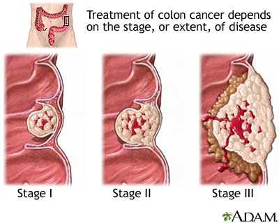
Most colorectal cancers follow the adenoma–carcinoma sequence:

A benign polyp forms first; over time, genetic mutations accumulate within it, and some polyps gradually become malignant. This process typically takes several years, which is why removing polyps during colonoscopy is both preventive and essential.

Clinical Presentation (What symptoms might patients report?)

Warning signs that should not be ignored include:

  • Bright red or dark blood in the stool, or a new and persistent change in bowel habits (constipation or diarrhea lasting more than 2–3 weeks)
  • Unexplained weight loss or fatigue caused by anemia
  • Pain or a sense of a mass in the lower abdomen or rectal area
  • Obstructive symptoms such as severe bloating, nausea, vomiting, and inability to pass stool or gas
  • In low-lying tumors: narrowing of the stool caliber or a sensation of incomplete evacuation

Any persistent symptom warrants medical evaluation — even in younger individuals.

Diagnosis

Colonoscopy + Biopsy

  • What it does: A camera is used to visualize the inside of the colon; polyps can be removed, and tissue samples can be taken from any suspicious mass.
  • Preparation: Complete bowel cleansing with prescribed solutions is required prior to the procedure. This step is essential for optimal visibility
  • Importance: Colonoscopy is both diagnostic and preventive, because polyps can be removed during the same procedure.

Pelvic MRI — Essential for Rectal Cancer

  • Why it matters: Pelvic MRI provides crucial information about: Depth of tumor invasion into the rectal wall (T stage) Tumor proximity to or involvement of the circumferential resection margin (CRM) Presence of extramural vascular invasion (EMVI) Suspicious regional lymph nodes

  • These findings determine whether preoperative chemoradiotherapy is needed and guide surgical planning. The MRI report should clearly specify: Distance to CRM EMVI status Tumor distance from the anal verge

Endorectal Ultrasound (ERUS / Endorectal Ultrasound)

  • Use: ERUS is helpful for very early tumors (e.g., T1–T2) when local excision is being considered. It provides detailed assessment of depth of invasion. It is also useful when MRI is unavailable or equivocal

CT Scan of Chest/Abdomen/Pelvis

  • Use: Required for systemic staging and detecting metastases, especially in the liver or lungs. If liver lesions are identified, further assessment begins to determine their resectability or potential for local control.

PET-CT and Molecular Testing

  • PET-CT:
    Used in selected cases, such as when findings are unclear or to assess treatment response. Molecular analysis: Pathology samples are typically tested for: MSI/MMR statusKRAS/BRAF mutationsOther relevant markersThese results influence the selection of targeted therapies and immunotherapy.

Treatment of Rectal Cancer

Treatment decisions in rectal cancer are complex and must be individualized. Each patient receives a coordinated plan developed by a multidisciplinary team, typically including a colorectal surgeon, a radiation oncologist, and a medical oncologist.

General Principles

  • If the tumor is considered threatening the resection margin on MRI, or if it is T3–T4 or associated with suspicious lymph nodes, neoadjuvant therapy (treatment before surgery) is typically recommended to shrink the tumor and increase the likelihood of achieving clear surgical margins.
  • If the tumor is very superficial and anatomically suitable, local excision through the anus may be an option in selected cases.

Neoadjuvant Therapy: Why and How?

There are two commonly used approaches:

  • Long-course chemoradiation, typically about 50.4 Gy combined with oral or intravenous chemotherapy. This regimen is used to shrink the tumor and increase the likelihood of sphincter preservation.
  • Short-course radiotherapy (5×5 Gy) followed by an interval before surgery. The choice between these approaches depends on tumor characteristics and institutional protocols.

The purpose of neoadjuvant therapy is to reduce tumor size, lower the risk of local recurrence, and enable surgery with clear margins.In some cases, newer protocols known as Total Neoadjuvant Therapy (TNT) incorporate systemic chemotherapy before surgery to enhance rates of complete response. These approaches are used in specialized centers with careful patient selection.

Standard Surgery: Total Mesorectal Excision (TME)

  • TME involves removing the tumor together with the mesorectum—the surrounding fatty tissue and lymph nodes—as an intact envelope. This technique is the gold standard for minimizing local recurrence.
  • Surgical options depend on tumor location:Higher rectal tumors may be treated with sphincter-preserving surgery and creation of a colorectal anastomosis.Very low tumors, when sphincter preservation is not feasible, may require complete removal of the rectum and anus with creation of a permanent stoma (abdominoperineal resection — APR).
  • When the anastomosis is constructed very close to the anus, a temporary diverting ileostomy is typically created to protect against leaks during early healing and is closed later.

Local Excision (TEM / TAMIS / TEO)

  • For large polyps or very early cancers (highly selected cases), local transanal excision using specialized instruments may be performed:
  • TEM (Transanal Endoscopic Microsurgery): Provides magnified visualization and precise instruments for tumor removal.
  • TAMIS (Transanal Minimally Invasive Surgery): A more modern technique using laparoscopic ports and standard instruments.
  • TEO (Transanal Endoscopic Operation): Similar to TEM but with different platforms.
  • These approaches preserve the rectum and allow rapid return to normal life, but they are appropriate only for strictly selected cases after accurate staging (MRI, ERUS, pathology).If deeper invasion or suspicious lymph nodes are present, local excision is insufficient.

Laparoscopic and Robotic Surgery

  • In experienced hands, laparoscopic and robotic approaches achieve oncologic outcomes comparable to open surgery. Potential advantages include:Smaller incisionsLess painFaster recoveryShorter hospital stayEnhanced visualization and greater instrument precision, especially valuable in the confined pelvic spaceA critical principle in these techniques is preservation of the pelvic autonomic nerves to reduce the risk of urinary and sexual dysfunction. Robotic systems with high magnification assist in nerve preservation and precise dissection. Long-term outcomes depend heavily on surgical expertise and patient selection.

TaTME (Transanal Total Mesorectal Excision)

  • In some highly specialized centers, TaTME is performed, allowing dissection from below (via the anus) upward.This approach can be beneficial for low tumors or patients with a narrow pelvis.
    Because of its technical demands, it should only be performed in expert centers.

Management of Metastases (Especially Liver Metastases)

  • The presence of metastases does not necessarily preclude curative treatment.When all liver lesions can be removed or locally controlled (surgical resection or ablation), and adequate liver volume remains afterward (usually at least 25–30% in a healthy liver), metastasectomy can be pursued with curative intent. Often, initial chemotherapy is given to shrink (downsize) the lesions. Subsequently, a multidisciplinary team—including a liver surgeon, colorectal surgeon, and oncologist—determines the treatment plan. Depending on the patient and institution, rectal surgery and liver metastasis surgery may be performed either simultaneously or in staged procedures.

Targeted Therapies and Immunotherapy

  • Based on molecular pathology results (MSI-high, MMR deficiency, KRAS/BRAF mutations), targeted agents or immunotherapy may be used in the metastatic setting or incorporated into TNT protocols.These decisions are highly individualized and depend on molecular test results.

The “Watch-and-Wait” Strategy

  • If a clinical complete response is observed after neoadjuvant therapy, some experienced centers may offer a watch-and-wait approach instead of immediate surgery. This requires extremely strict follow-up with repeated MRI scans and endoscopic evaluations. The strategy is reserved for carefully selected patients and should only be implemented in specialized centers.

Role of the Colorectal Surgeon

  • The colorectal surgeon plays a central role in decision-making: interpreting the MRI findings, choosing between neoadjuvant therapy and upfront surgery, determining whether sphincter preservation is possible or if APR is required, and coordinating multidisciplinary management of metastases.
  • Rectal surgery—particularly TME and the associated reconstructive procedures—requires specialized skill and experience. Nerve preservation and the maintenance of urinary and sexual function depend heavily on the surgeon’s expertise.
  • Any major intervention—especially reconstructive operations or advanced techniques such as TaTME or metastasectomy—should be planned within a multidisciplinary team with active involvement of the colorectal surgeon.

پیگیری پس از درمان

  • Year 1: Clinical visits and blood tests (CEA) every 3 months; imaging (CT or MRI) every 6–12 months depending on disease stage.
  • Year 2: Clinical visits and CEA testing every 3–6 months; CT scan every 6–12 months.
  • Years 3–5: Clinical visits and CEA every 6 months; imaging every 6–12 months based on individual risk.
  • Colonoscopy: Typically performed one year after surgery, and then at intervals determined by findings (for example at 1, 3, and 5 years, or as recommended by the care team).
  • After 5 years: If the patient remains stable, follow-up intervals may be extended, but annual evaluations should continue.

Frequently Asked Questions About Rectal Cancer

At what age should I begin screening?

For individuals at average risk, screening should begin at age 45.
If you have a family history of colorectal cancer or a genetic syndrome, screening typically starts 10 years before the youngest affected family member’s age at diagnosis, or at age 40—whichever comes first.
Consult a gastroenterologist or a genetic counseling center for personalized guidance.

The report should specify the distance from the tumor to the circumferential resection margin (CRM), the presence or absence of EMVI, the T stage, the tumor’s distance from the anal verge, and any suspicious lymph nodes. These findings are essential for determining the appropriate treatment plan.

It refers to chemoradiation given before surgery to shrink the tumor and increase the likelihood of sphincter preservation or achieving clear surgical margins.
Neoadjuvant therapy is typically used for locally advanced tumors or those that threaten the circumferential resection margin (CRM).

No. If all liver lesions can be surgically removed or locally controlled, and the patient’s overall condition is suitable, metastasectomy can be performed with curative intent. This decision is made by a multidisciplinary team.

Both techniques provide excellent oncologic outcomes in well-equipped centers. Robotic and laparoscopic approaches offer advantages in the narrow pelvic space, allowing for more precise dissection and better nerve preservation.
The choice depends on the surgical team’s experience and the patient’s specific circumstances.

Summary for Patients

Rectal cancer is a condition that, in many cases, is treatable or manageable when detected early. Diagnosis is based on colonoscopy and pelvic MRI, and treatment decisions typically involve a combination of chemoradiation and surgery using the TME technique.

In modern centers, laparoscopic or robotic approaches—and, in selected cases, local excision techniques such as TEM, TAMIS, or TEO—can offer effective treatment with better functional outcomes and fewer complications. However, all of these options require an experienced team and careful patient selection.

Any major intervention—especially complex surgery—should be guided by a specialized colorectal surgeon and managed within a multidisciplinary team.