Anal Fistula

Introduction

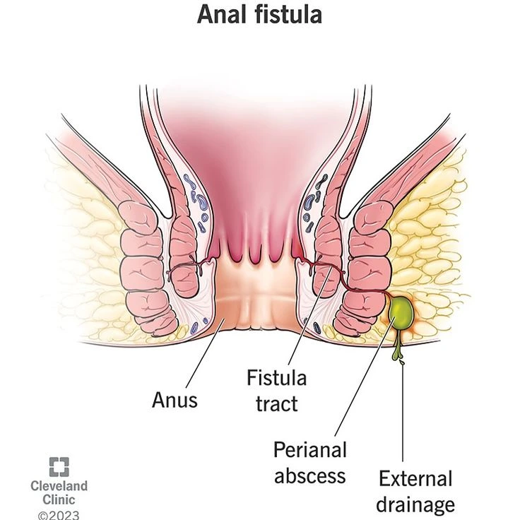
An anal fistula is an abnormal tract or tunnel that forms between the internal space of the anal canal—typically originating from a cryptoglandular site or following an anorectal abscess—and the perianal skin. In simple terms, it is a “purulent pathway” that opens from inside the rectum to the outside, and is usually associated with recurrent discharge, pain, or perianal irritation. The goal of treatment is to close this tract while preserving sphincter function.

Anatomy

To understand anal fistulas, three key components must be recognized:
  1. The sphincters
  2. Anatomic spaces and potential pathways
  3. The internal and external openings
  1. The Sphincters

  • Internal sphincter: A circular smooth muscle that functions involuntarily and is responsible for maintaining the baseline anal tone.
    External sphincter: A striated, voluntary muscle that provides conscious control over continence.

Proper sphincter function determines the risk of postoperative incontinence; the less muscle divided, the lower the likelihood of dysfunction.

  1. Spaces and Pathways

  • The anal crypts and their associated cryptoglandular structures at the base of the crypts are the most common origin of fistulas.
  • Fistulas are classified based on their course in relation to the sphincter muscles: intersphincteric, transsphincteric, suprasphincteric, and extrasphincteric. This classification guides the choice of treatment and predicts the risk of incontinence.
  1. Internal and External Openings

  • Internal opening: The site where the fistula communicates with the anal canal, usually at the internal end of a crypt.
  • External opening: The cutaneous opening through which purulent discharge exits. Accurate identification of both openings is essential for successful treatment.

Types of Anal Fistulas

An anal fistula is a pathological tract or tunnel between the internal space of the anal canal and the perianal skin, typically developing after an anorectal abscess.

Types Based on Their Course Relative to the Sphincter Complex:

  • Intersphincteric: The most common type.
  • Transsphincteric: Traverses both the internal and external sphincters.
  • Suprasphincteric: Passes above the internal sphincter.
  • Extrasphincteric: Rare; often associated with external disease processes or trauma.

Etiology (Causes and Pathophysiology)

  • Anal fistulas are most commonly observed in middle-aged adults, with a slightly higher prevalence in men than in women.
  • The most frequent underlying condition is a history of anorectal abscess that has progressed to fistula formation.
  • Additional factors that increase the risk or complexity of fistulas include inflammatory bowel disease (particularly Crohn’s disease), poorly controlled diabetes, prior anorectal surgery, pelvic trauma or radiotherapy, and infectious or immunocompromised states such as HIV.
  • In Crohn’s disease, fistulas tend to be more complex, branching, and treatment-resistant, often requiring combined medical therapy and staged surgical intervention.

Prevention of Anal Fistula

  • Timely treatment and drainage of anorectal abscesses is the most important preventive measure; proper abscess drainage prevents progression to fistula formation.
  • Control of underlying conditions such as Crohn’s disease and diabetes is essential.
  • Maintaining local hygiene and managing constipation help reduce pressure and irritation in the area
    • In the presence of fever or signs of an abscess, prompt medical evaluation is crucial; delay is the most common factor leading to fistula development.

Pathogenesis (Mechanism of Disease Development)

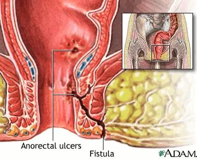
The typical sequence begins with obstruction or infection of a crypt gland, leading to the formation of a localized abscess. If the abscess is not adequately drained, or if purulent material extends along areas of tissue weakness, a tunnel-like tract may persist after drainage, maintaining communication between the internal mucosa and the external skin—this persistent tract is the fistula.
In Crohn’s disease, the inflammatory process within the bowel wall can directly generate fistulas without a clearly defined preceding superficial abscess.

Clinical Manifestations

  • Persistent or intermittent discharge of purulent or foul-smelling fluid from a cutaneous opening near the anus.
  • Mild to moderate pain, with occasional sharp or episodic pain if the tract becomes temporarily obstructed.
  • Perianal irritation or dermatitis (contact eczema) around the external opening due to ongoing discharge.
  • Fever and significant pain may occur if a secondary abscess or active infection is present.
  • In complex fistulas or those situated close to the sphincter complex, patients may report altered evacuation patterns or incontinence; in such cases, careful clinical examination is essential.

Diagnosis of Anal Fistula

A) History and Clinical Examination

  • History should focus on the pattern of discharge, prior anorectal abscesses, underlying conditions such as Crohn’s disease, and any history of surgery or traumatic childbirth.
  • Examination includes inspection of the perianal skin to identify the external opening, and digital rectal examination to detect signs of an internal opening or an indurated tract. In some patients, office examination may be painful and may need to be performed under local anesthesia or in the operating room.

B) Imaging Studies

  1. Fistula-mapping MRI (Pelvic MRI with a fistula protocol)
  • The best modality for accurately delineating fistulous tracts, side branches, the internal opening, and their relationship to the sphincter complex.
  • Technique: T2-weighted and fat-suppressed sequences are obtained, and in some cases gadolinium may be instilled through the external opening or into the anal canal to enhance visualization of the tract. The radiologist’s report should specify the exact course of the fistula, the location of the internal opening, and the presence of any occult abscesses or secondary branches.
  • Clinical use: Essential before sphincter-preserving procedures and for planning surgeries such as LIFT or advancement flap repair.
  1. Endoanal Ultrasound (EAUS)
  • A radial or linear probe is inserted into the anal canal to visualize the sphincter muscles and tracts close to the canal.
  • It is useful for detecting sphincter defects, internal openings near the canal, and relatively superficial fistulas. It is less expensive and faster than MRI, but its sensitivity is lower in complex fistulas.
  1. EUA (Examination Under Anesthesia) 
  • In many cases, accurate diagnosis involves probing the tract and injecting a dye (such as methylene blue or hydrogen peroxide) through the external opening to identify the internal opening. During the same session, a seton may be placed or drainage may be performed. This approach provides both diagnostic and therapeutic benefit.
  1. Sigmoidoscopy/Colonoscopy
  • Colonoscopy is indicated when there is evidence or a history of inflammatory bowel disease, gastrointestinal symptoms, or atypical fistulas. It is particularly important in cases with multiple or non-midline fistulas to exclude Crohn’s disease or other underlying pathology.
  1. Other Imaging Modalities
  • Perineal ultrasound or CT can be helpful in centers where MRI is not available; however, MRI remains the gold standard for fistula mapping and identification.

c) Additional Laboratory Tests

  • Blood tests may be performed to evaluate inflammatory markers and to assess glycemic control in patients with diabetes. If Crohn’s disease is suspected, further inflammatory studies should be ordered and the patient should be referred to a gastroenterologist.

Management — Principles, Techniques, Indications, and Outcomes

A key principle is that treatment aims to close the fistula tract while minimizing sphincter injury and preserving continence. The choice of technique depends on the type of fistula (simple or complex), the location of the internal opening, the condition of the sphincter complex, and the presence of underlying diseases such as Crohn’s disease.

A) Initial / Conservative Management

  • Control of infection is essential, and if an active abscess is present, partial drainage and placement of a seton for long-term drainage should be performed before any attempt at definitive closure.
  • In Crohn’s disease, medical therapy (including steroids, immunomodulators, and anti-TNF agents) is recommended prior to any sphincter-preserving procedure, as operating during periods of high inflammatory activity increases the risk of recurrence. Close collaboration with rheumatology and gastroenterology specialists is necessary.

B) Surgical and Sphincter-Preserving Techniques (Description, Indications, and Outcomes)

  1. Fistulotomy

  • Opening the fistula tract and allowing it to heal by secondary intention.
  • Indication: Simple fistulas involving less than approximately 30% of the external sphincter (the exact decision requires careful sphincter assessment).
  • Advantages: High success rates, reported as 80–95% in many series.
  • Disadvantages: If too much sphincter muscle is divided, there is a risk of incontinence; therefore, this technique is reserved for simple, superficial fistulas.
  1. Seton — Types and Applications

  • Loose (non-cutting) seton: A soft suture or silicone loop that keeps the tract open to allow continuous drainage and reduction of inflammation; appropriate for complex fistulas or for patients with active inflammation, such as those with Crohn’s disease. Its primary purpose is long-term drainage while protecting the sphincter.
  • Cutting seton: Applies gradual tension to the tract, producing controlled division of the sphincter with subsequent healing. Due to the risk of incontinence, its use has become more limited in modern practice.
  • Note: Seton placement is often the first step in managing complex fistulas to control inflammation before definitive treatment.
  1. LIFT (ligation of intersphincteric fistula tract)

  • The principle of the procedure is to identify and ligate the fistula tract within the intersphincteric space while dividing only the intersphincteric portion, without cutting the external sphincter.
  • Indication: Low to mid-transsphincteric fistulas in which the intersphincteric portion of the tract is accessible.
  • Advantages: Sphincter preservation with a low risk of incontinence; initial success rates in studies range from approximately 60–80%.
  • Limitations: May be insufficient in highly complex or branching fistulas.
  1. Advancement flap 

  • This technique involves excising the fibrotic tissue around the internal opening and covering the site with a healthy mucosal or cutaneous flap to achieve closure.
  • Indication: Fistulas with a large internal opening, cases in which sphincter division is contraindicated, or as a salvage option after failure of other techniques.
  • Advantages: Preservation of sphincter integrity; success rates reported around 50–70% in various series.
  • Disadvantages: Requires meticulous surgical technique, and recurrence remains possible.
  1. (Fistula plug / Fibrin glue)

  • This method involves filling the fistula tract with fibrin glue or biological plugs (such as porcine collagen) to achieve internal closure of the pathway.
  • Indication: Selected fistulas that are short and have minimal branching, or in patients who prefer to avoid open surgical procedures.
  • Advantages: Minimally invasive and sphincter-preserving.
  • Disadvantages: Variable success rates (approximately 40–60%), and repeat procedures or alternative surgery may be required.
  1. VAAFT, FiLaC, Laser closure

  • VAAFT (Video-Assisted Anal Fistula Treatment): A camera is introduced into the fistula tract to allow direct visualization, internal debridement, and subsequent closure of the internal opening.
  • FiLaC (Fistula Laser Closure): A laser probe is used within the tract to thermally ablate the fistula from the inside, promoting collapse and scarring of the pathway.
  • Indication: Well-defined fistulas in specialized centers, particularly for patients who prefer less invasive options.
  • Advantages: Sphincter preservation and a shorter recovery period.
  • Disadvantages: Long-term evidence remains limited, and reported success rates vary between 50–80%.
  1. Combined Repairs and Complex Surgical Approaches

  • In recurrent fistulas or those with horseshoe or supralevator extension, combined techniques, staged procedures, or collaboration with pelvic or colorectal surgical specialists may be required.

۸.۳ Selection of Treatment Method

  • Simple, superficial fistulas → Fistulotomy (provided that sphincter involvement is minimal).
  • Complex fistulas or those with substantial sphincter involvement → Initial drainage with a seton followed by a sphincter-preserving procedure such as LIFT, advancement flap, or endoscopic techniques.
  • In Crohn’s disease → Primary medical therapy along with seton placement for drainage; final management should be determined by a multidisciplinary team.
  • The overarching goal is always to close the internal opening while preserving maximal sphincter function.

Outcomes

(Approximate ranges based on pooled data from review studies; presented as general estimates.)

  • Fistulotomy for simple fistulas: success rates above 80–90%.
  • LIFT: 60–80% success in various reports, with a low risk of incontinence.
  • Advancement flap: approximately 50–70% success; technically more demanding.
  • Plug or fibrin glue: variable outcomes, generally 40–60%; suitable for selected cases.
  • VAAFT / FiLaC: promising short-term results, though long-term data remain limited.

Factors associated with treatment failure or recurrence include complex or branching fistulas, a history of Crohn’s disease, active infection, incomplete identification of the internal opening, and inadequate management of the sphincteric component.

Role of the Colorectal Surgeon

  • Comprehensive preoperative evaluation using MRI and/or EAUS, with treatment decisions tailored to fistula type and sphincter function.
  • Performance of sphincter-preserving techniques (such as LIFT, advancement flap, and endoscopic procedures) when indicated.
  • Multidisciplinary collaboration with gastroenterologists and other specialists in patients with complex fistulas or Crohn’s disease.
  • Patient education regarding postoperative care, the possibility of recurrence, and the need for structured follow-up.

Frequently Asked Questions About Anal Fissure

Does an anal fistula always require surgery?

In most cases, yes. Persistent discharge and ongoing infection increase the risk of complications, including incontinence. In patients with Crohn’s disease or certain special conditions, initial medical therapy or placement of a seton for drainage may be sufficient before considering definitive surgery.

Incontinence may occur if an inappropriate technique is chosen or if an excessive portion of the sphincter complex is divided. For this reason, sphincter-preserving procedures and management by a colorectal surgeon are essential in complex fistulas.

Depending on the degree of inflammation and the presence of a seton, a waiting period of several weeks to a few months is typically required to allow inflammation to subside and to optimize conditions for definitive repair.

They can be managed, but treatment often requires a combination of medical therapy (such as anti-TNF agents) and staged surgical procedures, and recurrence is more common than in non-Crohn’s fistulas.

Fistula-mapping MRI is the most accurate method for delineating fistula tracts and planning surgical management.

Patient Summary

An anal fistula is an abnormal tunnel connecting the inside of the anal canal to the surrounding skin, typically developing after an anorectal abscess. Its main symptom is recurrent drainage of pus or bloody fluid. Diagnosis relies on clinical examination and detailed imaging, particularly MRI.
The goal of treatment is to close the tract while preserving continence. Fistulotomy is effective for simple fistulas, whereas sphincter-preserving techniques (such as LIFT, advancement flap, seton placement, or endoscopic approaches) are preferred for complex cases.
Patients with Crohn’s disease or other underlying conditions also require concurrent medical therapy. Collaboration with a colorectal surgeon and a multidisciplinary team improves outcomes and enhances the likelihood of successful treatment.