Anal Fissure

فیشر شقاق مقعدی

Introduction

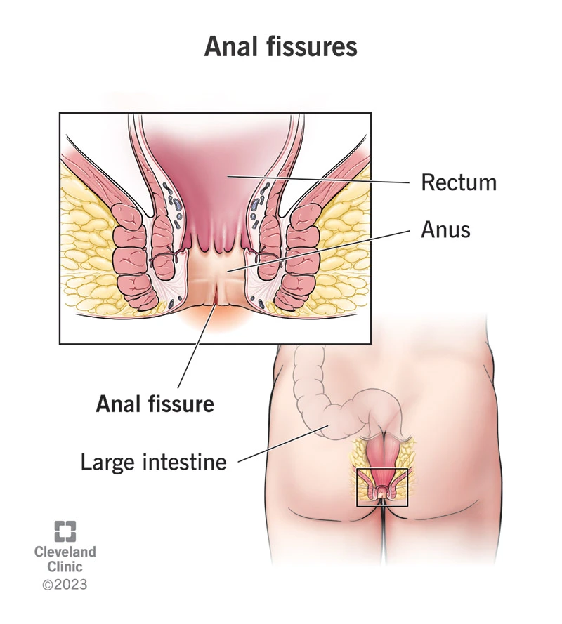
An anal fissure is a longitudinal tear in the delicate skin lining the anal canal (the anoderm).
This tear causes sharp pain during bowel movements and bright red bleeding.
A fissure may be acute (recent and short-term) or chronic (lasting more than 6–8 weeks, often with fibrotic edges or a sentinel skin tag).

Anatomy

To better understand anal fissures, it is helpful to be familiar with a few key anatomical structures:
  • Anoderm: The thin, highly pain-sensitive skin lining the anal canal.
  • Dentate line: The boundary between the upper mucosal lining and the lower pain-sensitive skin.
  • Internal anal sphincter: A ring of involuntary smooth muscle that provides the baseline resting tone of the anal canal; this muscle typically maintains relatively high resting pressure.
  • External anal sphincter: A voluntary skeletal muscle that allows conscious control over bowel movements.
  • Conjoined longitudinal muscle: A layer located between the mucosa and the deeper muscular structures, contributing to the firmness and structural stability of the anal canal.

Types of Anal Fissures

  • Acute fissure: A recent tear in the anoderm, typically presenting with sudden-onset pain and bleeding.
  • Chronic fissure: A tear persisting for more than 6–8 weeks, often accompanied by fibrotic edges, a sentinel pile (a protective skin tag), or an enlarged internal papilla.
  • Off-midline or multiple fissures: Fissures located in atypical positions or occurring in multiple sites warrant evaluation for underlying conditions such as inflammatory bowel disease (Crohn’s), infections, or rare lesions.

Epidemiology (Prevalence, Patterns, and Who Is Most Affected?)

  • Anal fissures are most common in young to middle-aged adults (approximately 20–45 years).
  • In 70–90% of cases, the fissure is located in the posterior midline; in women—especially after childbirth—anterior fissures are also more likely.
  • The presence of off-midline, multiple, or treatment-resistant fissures is a warning sign for potential underlying conditions and warrants further evaluation.
آناتومي مقعد

Etiology (Causes and Pathophysiology)

Several factors work together to create a cycle that prevents proper healing:

  1. Mechanical trauma: Passage of hard stool, excessive straining, or recurrent diarrhea can tear the anoderm.
  2. Internal sphincter spasm: Elevated resting pressure in the anal canal reduces blood flow to the base of the fissure.
  3. Local ischemia and impaired healing: Diminished blood supply combined with sphincter spasm delays healing and promotes progression to a chronic fissure.

The result of this cycle is: pain → sphincter spasm → reduced blood flow → impaired healing → worsening pain.

Clinical Manifestations (What Symptoms Do Patients Report?)

  • Sharp, fleeting pain during bowel movements, which may persist for several minutes to several hours afterward.
  • Bright red blood appearing as streaks on toilet paper or on the surface of the stool.
  • Fear of defecation, resulting in secondary constipation, avoidance of using the toilet, itching, or burning.
  • In chronic cases: the presence of an external sentinel pile or a white-based ulcer at the site of the fissure.

If the pain is too severe and the patient is fearful of examination, the evaluation can be performed under local anesthesia.

Diagnosis

A) Detailed Medical History

Key questions include: the duration and severity of pain, bowel movement patterns, presence of constipation or diarrhea, pregnancy or childbirth history, use of anticoagulant medications, and any warning signs such as weight loss, fever, significant bleeding, or systemic symptoms. This information is essential for determining whether further evaluation is required.

B) Clinical and Visual Examination

A careful inspection of the perianal skin usually confirms the diagnosis; fissures are most commonly found in the posterior midline. Digital rectal examination (DRE) is performed only if the patient can tolerate it or if it is done under local anesthesia.

C) Anoscopy / Proctoscopy

After initial pain has subsided, simple anoscopy helps visualize the exact location of the fissure, as well as any associated papillae or hemorrhoids. Proctoscopy is used to examine higher sections of the rectal canal.

D) When Is Colonoscopy Necessary?

If a fissure is off-midline, multiple, treatment-resistant, or accompanied by warning signs—such as unexplained weight loss or long-standing changes in bowel habits—colonoscopy is necessary to rule out inflammatory bowel disease or more proximal lesions. This distinction is important because not all fissures require colonoscopy, but in atypical cases or in patients over the age of 40, a full colorectal evaluation is recommended.

E) Anorectal Manometry (In Select Cases)

In patients who are candidates for treatments that alter sphincter tone (such as Botox injection), or when evaluating the cause of preoperative incontinence, anorectal manometry provides measurements of resting and squeeze pressures of the sphincters and helps guide clinical decision-making.

Treatment of Anal Fissure

(Step-by-step guide from conservative management to surgical options)

Principle:Management begins with the least invasive and simplest measures. If adequate healing does not occur after a reasonable period (typically 6–8 weeks), or if the fissure becomes chronic, treatment progresses to the next tier of options.

درمان بواسیر با حمام آبگرم

A — First-line Measures (Recommended for All Patients)

  • Increasing dietary fiber intake (approximately 20–30 grams per day) along with adequate hydration.
  • Using stool softeners (such as polyethylene glycol) to keep the stool soft and prevent hardness.
  • Taking warm sitz baths for 10–15 minutes, several times a day and after bowel movements.
  • Correcting toileting habits: avoiding straining and minimizing prolonged sitting on the toilet.

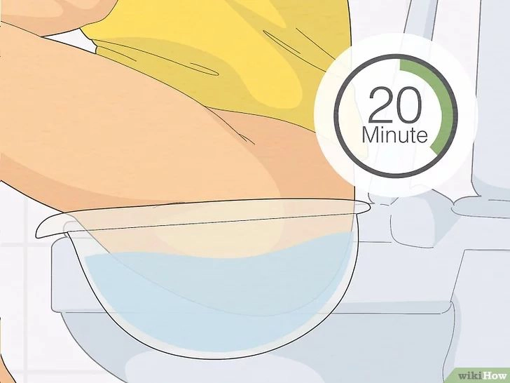
These simple measures lead to improvement in many patients during the acute phase; however, the longer the fissure has been present, the lower the likelihood that it will heal with conservative measures alone.

B — Topical Medications to Reduce Sphincter Tone (First-line Medical Therapy)

  • Nitroglycerin (GTN) ointment: Relaxes the internal anal sphincter, increases local blood flow, and promotes healing. The most common side effect is headache, which is usually transient. Treatment typically continues for several weeks.
  • Topical calcium-channel blockers (diltiazem or nifedipine): These agents have an effect similar to GTN by relaxing the internal sphincter and improving local blood flow, but they are associated with fewer headaches and are often better tolerated.

Studies show that these medications are effective for acute fissures and for some chronic cases; however, their healing rates are lower compared with surgical treatment.

C — Botulinum Toxin Injection — “Next Step or a Pre-surgical Alternative”

  • Rationale: Botulinum toxin induces a temporary, partial paralysis of the internal anal sphincter, reducing resting tone and allowing the fissure to heal.
  • How it is performed: The injection is administered in the office or operating room by placing the toxin into two to three sites within or adjacent to the internal anal sphincter.
    It typically requires only local anesthesia and is performed as an outpatient procedure.
  • Success rate: Systematic reviews report healing rates of approximately 60–77%, depending on the study and patient population. However, a notable proportion of patients experience recurrence over the long term and may require repeat injections or surgical intervention. Major complications are rare, though temporary leakage of gas or minor discharge may occur.

D — Surgical Treatment: Lateral Internal Sphincterotomy (LIS)

The surgical gold standard

  • What it is and why it is used: This procedure involves making a small lateral incision in the internal anal sphincter to reduce resting tone, thereby improving blood flow and allowing the fissure to heal. It offers the highest healing rates and the lowest recurrence rates among available treatments.
  • Types: The procedure may be performed using an open technique (direct incision) or a closed technique (using a guided subcutaneous approach). Both methods are effective, and the choice largely depends on the surgeon’s experience and preference.
  • Outcomes: Long-term healing rates are generally high, with multiple studies reporting success rates of approximately 90–95%. However, reducing sphincter tone may, in rare cases, lead to some degree of incontinence. According to reputable sources, persistent incontinence is uncommon, but reported rates vary, with some series noting a small percentage of cases.
    Careful patient selection and assessment of risk factors—such as a history of prior sphincter injury—are therefore essential.

E — Alternative Surgical Techniques

(For patients at risk of incontinence or in whom LIS is not appropriate)

  • Mucosal or anocutaneous advancement flap: This technique involves excising the fibrotic tissue and covering the fissure with a healthy flap. It is particularly suitable for patients with low sphincter tone or those at increased risk of incontinence, serving as an alternative to LIS when sphincter-sparing is essential.
  • Combination or tailored techniques: Customized approaches may be used depending on the clinical examination, childbirth history, or any prior sphincter injury. These individualized strategies are selected to optimize healing while preserving sphincter function.

When Is Surgery Necessary? (Clear Indications)

  • Symptomatic chronic fissure that does not respond to medical therapy (typically after 6–8 weeks of appropriate treatment).
  • Persistent pain or significant impairment in quality of life.
  • Recurrent fissures despite medical management or prior Botox injections.
  • Patients in whom Botox has failed or who prefer a more definitive solution—provided they fully understand the benefits and potential risks.

Complications and Safety Considerations (What Patients Should Know)

  • After medical therapy: Headache (with GTN), local discomfort, and the possibility of recurrence.
  • After Botox injection: Temporary leakage of gas or minor discharge may occur in some patients.
  • After LIS: Postoperative pain, bleeding, and infrequent infections may occur. The risk of incontinence is variable across reports — most patients experience only temporary difficulty controlling gas or minor moisture, while persistent incontinence is uncommon and seen in a small percentage of cases. Careful patient selection, consideration of childbirth history or prior sphincter injury, and preoperative assessment with endoanal ultrasound or manometry help minimize this risk.

Role of the Colorectal Surgeon

  • Accurate diagnosis, including distinguishing a simple fissure from one caused by an underlying condition.
  • Selecting a logical, stepwise treatment pathway (from conservative therapy → Botox → surgery) and performing interventional procedures with preservation of sphincter function as a priority.
  • Conducting or coordinating specialized evaluations (such as endoanal ultrasound [EAUS] and anorectal manometry) in complex or suspicious cases, and determining whether sphincter-sparing techniques or advancement flap procedures are appropriate.

Frequently Asked Questions (FAQs)

Does an anal fissure always require surgery?

No — many acute fissures heal with dietary fiber, stool softeners, and sitz baths. Only chronic or treatment-resistant fissures require more advanced or invasive interventions.

Botox is a temporary, minimally invasive option with a moderate success rate. LIS offers a higher and more durable healing rate, but carries a slightly greater risk of functional side effects. The choice between the two depends on clinical factors and the patient’s preferences.

Persistent incontinence is not common, but reported rates vary across studies. Pre-operative risk assessment — including childbirth history, endoanal ultrasound (EAUS), and anorectal manometry — is essential for minimizing the likelihood of postoperative functional problems.

Summary for patients

An anal fissure is a painful tear in the skin of the anal canal, typically accompanied by pain and bright red bleeding. Most fissures improve with dietary changes, stool softeners, and warm sitz baths. If healing does not occur within 6–8 weeks, treatments such as topical medications, Botox injections, or a minor surgical procedure (lateral internal sphincterotomy) may be recommended. The majority of patients recover well with appropriate therapy.Choosing the most suitable treatment requires a thorough examination and consultation with a colorectal surgeon.