Descending Colon Cancer

كولونوسكوپي

Introduction

Descending colon cancer is a tumor that begins in the inner lining of the large intestine in the descending segment (on the left side of the body). It may initially grow without noticeable pain or symptoms, then infiltrate deeper layers of the bowel wall, spread to nearby lymph nodes, or metastasize through the bloodstream to other organs such as the liver or lungs.

Epidemiology (Prevalence and Risk Factors)

Compared with ascending and sigmoid colon cancers, descending colon cancer is less common and accounts for about 5–7% of all colorectal cancer cases. Globally, this cancer typically appears in the sixth and seventh decades of life and is often associated with symptoms such as changes in bowel habits and overt bleeding. In Iran, official statistics rarely report cases by exact location, but available evidence suggests that descending colon cancer has a distribution similar to global patterns and, unlike right-sided colon cancers, is sometimes reported at relatively younger ages.

Pathogenesis (Mechanism of Disease Development)

This process is usually long and stepwise, and understanding it is essential for appreciating the importance of screening:

  1. Initiation — Polyp formation (adenomatous polyp): A benign protrusion may form in the mucosal layer of the bowel, known as a polyp. Not all polyps are dangerous, but some are adenomatous and, if not removed, may undergo malignant changes over several years.
  2. Accumulation of genetic mutations: Over time, some of these polyps develop mutations in genes that regulate cell division and DNA repair. These mutations gradually disrupt the normal control of cellular growth.
  3. Progression to an invasive tumor: Once enough mutations have accumulated, the polyp can transform into an invasive tumor that penetrates the mucosa and extends into the muscular and outer layers of the colon wall.
  4. Spread: Some cancer cells may enter lymphatic or blood vessels and seed themselves in lymph nodes or distant organs—most commonly the liver, and subsequently the lungs.

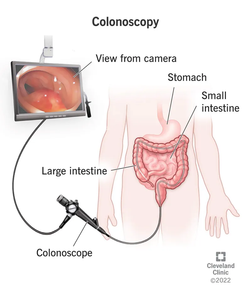
Key point: This sequence typically takes many years; therefore, regular colonoscopy and polyp removal greatly increase the chance of preventing cancer.

Etiology (Causes and Disease Mechanisms)

I divide the risk factors into two groups: non-modifiable and modifiable, and explain the role of each.

Non-modifiable Factors

  • Age: Most cases occur after age 50, although younger cases do occur.
  • Family history and genetics: Having a first-degree relative with colorectal cancer, or a hereditary syndrome such as Lynch syndrome or FAP, increases the risk. Earlier and more frequent screening is required in these situations.
  • History of polyps or long-standing intestinal inflammation (such as ulcerative colitis), which creates a background that promotes malignant transformation.

Modifiable Factors

  • Low-fiber diet and processed meats: Diets low in fiber lead to harder stools and straining; this alters intracolonic pressure and creates a setting that favors polyp formation. In contrast, fiber (fruits, vegetables, whole grains) softens the stool and has a protective role.
  • Obesity and physical inactivity: Obesity and a sedentary lifestyle are associated with chronic inflammation and metabolic changes that increase cancer risk. Regular physical activity and modest weight reduction can help lower the risk.
  • Smoking and heavy alcohol use: Both factors are linked to a higher risk of gastrointestinal cancers. Quitting smoking and reducing alcohol intake are beneficial.
  • Long-term use of NSAIDs and certain medications: Nonsteroidal anti-inflammatory drugs can alter bleeding risk or other gastrointestinal effects. Some studies suggest complex interactions between NSAID use and cancer risk; stopping or continuing these medications should be done in consultation with a physician.
  • Antioxidant-rich diets and high intake of fruits/vegetables: Diets rich in plant-based foods, antioxidants, fruits, and vegetables are associated with a reduced risk.

Clinical Manifestations

  • A new and persistent change in bowel habits (diarrhea or constipation lasting more than 2–3 weeks).
  • Blood in the stool or occult bleeding leading to anemia (fatigue, pallor).
  • Unexplained weight loss and loss of appetite.
  • Persistent pain or a palpable mass in the left side of the abdomen.
  • Signs of bowel obstruction (severe bloating, nausea, inability to pass gas or stool).

Some early symptoms may be mild or nonspecific; however, the presence of anemia or weight loss should always be taken seriously and warrants thorough evaluation.

Diagnosis

  • Colonoscopy with biopsy: The most important and definitive diagnostic method. The entire colon is inspected with a camera, and if a lesion is detected, a tissue sample is taken so pathology can determine the type of tumor and its degree of invasiveness.
  • Blood tests: Assessment for anemia (CBC), evaluation of liver function, and measurement of CEA (a tumor marker). CEA is useful for follow-up but is not diagnostic on its own.
  • Staging imaging (contrast-enhanced CT of the abdomen and pelvis, and chest CT): Used to identify local tumor extension, lymph node involvement, and metastases to the liver or lungs.
  • Molecular pathology evaluation: The biopsy may be tested for MSI/MMR status or mutations such as KRAS/BRAF. This information helps guide the selection of targeted therapies and immunotherapy.

Treatment

Treatment is determined by several factors: the stage of the tumor (its size, depth of invasion, and lymph node status), the patient’s overall condition, and the presence or absence of metastasis.

A — Surgery

  • In most cases, the procedure involves removing the descending portion of the colon (left colectomy or segmental colectomy). The surgeon removes the tumor along with the regional lymph nodes to allow accurate staging.
  • If the situation is an emergency (such as obstruction or perforation), the surgery may be performed in stages, and a temporary stoma may be created.
  • The operation can be done laparoscopically (minimally invasive) or as an open surgery; in well-equipped centers, laparoscopy is typically associated with less postoperative pain and a faster return to normal activities.

B — Adjuvant and Neoadjuvant Chemotherapy

  • Adjuvant therapy (after surgery): If lymph nodes are involved or high-risk features are present, chemotherapy is recommended after surgery to reduce the likelihood of recurrence.
  • Neoadjuvant therapy (before surgery): In selected cases, it may be used to shrink the tumor or achieve faster disease control, although its use in colon cancer is less common than in rectal cancer.

C — When the Cancer Has Spread to the Liver — Can Liver Metastases Be Surgically Removed?

Goal: If all liver lesions can be completely removed or locally controlled, and the remaining liver volume after resection is sufficient, a curative treatment approach may be possible. Key considerations include:

  1. Number and size of liver lesions: Single or few lesions located in surgically accessible areas are usually better candidates. The main point is that all lesions must be removable or controllable.
  2. Location of lesions and future liver remnant (FLR): After resection, the remaining liver volume must be adequate for normal function (approximately 25–30% in a healthy liver; more is required if the liver is damaged).
  3. Response to systemic chemotherapy: Sometimes chemotherapy is given first to shrink the lesions (downsizing) to make surgical removal feasible.
  4. Presence or absence of extrahepatic disease: If disease is also present in the lungs or other organs, the overall situation must be evaluated. In select cases, both liver and lung metastases can be removed, either simultaneously or in staged procedures.
  5. Overall patient condition and liver function: The patient must be able to tolerate surgery, and the liver must not be severely impaired.

 Overall patient condition and liver function:The patient must be able to tolerate surgery, and the liver must not be severely impaired.

D — Targeted Therapies and Immunotherapy

  • Depending on the molecular characteristics of the tumor (such as MSI-high status or specific mutations), targeted therapies or immunotherapy may be effective. This decision is based on the results of molecular pathology testing.

E — Postoperative Follow-up

  • First year: Clinical visit every 3 months + blood tests (including CEA) every 3 months for early surveillance.
  • Year 2: Clinical visit every 3–6 months + CEA every 3–6 months.
  • Years 3–5: Clinical visit every 6 months, with CEA every 6 months; abdominal and chest CT scans are typically performed every 6–12 months depending on risk level and disease stage.
  • Postoperative colonoscopy: Usually performed one year after surgery. If normal, it is typically repeated after 3 years, and then every 5 years thereafter, depending on findings.

After 5 years: If everything remains stable, the intensity of follow-up decreases, but annual visits and general health monitoring continue.

Frequently Asked Questions About Descending Colon Cancer

Does diet really make a difference?

Yes. A high-fiber diet, reducing processed meats, maintaining a healthy weight, and staying physically active all help lower the risk and support recovery after treatment.

Not necessarily. In many cases, if the liver lesions are removable or can be locally controlled and the patient is in good overall condition, surgical removal or local treatment of liver metastases can still be pursued with curative intent. This decision is made by a multidisciplinary team.

Usually one year after the operation; if the result is normal, the next exam is done after 3 years, and then every 5 years. However, the schedule may be adjusted depending on previous findings.

There is no absolute guarantee that cancer will “never return,” but evidence shows that a healthy lifestyle reduces the risk of recurrence and improves quality of life.

High fever, sudden and severe abdominal pain, heavy rectal bleeding, persistent nausea and vomiting, or foul-smelling discharge from the surgical incision are warning signs that require urgent medical attention.

Summary for Patients

Descending colon cancer is a condition that can be diagnosed and, in many cases, effectively treated—especially when detected early. A combination of surgery, chemotherapy when needed, and careful follow-up greatly improves the chances of successful treatment. Positive changes in diet, weight, and physical activity can help reduce the risk of developing or recurring disease and support better treatment outcomes.