Uterine and Vaginal Prolapse

پرولاپس

Introduction

Uterine and vaginal prolapse refer to the descent of the uterus or vaginal walls into the vaginal canal due to weakened pelvic floor muscles and ligaments. These conditions can lead to symptoms like pelvic pressure, bulging, and sometimes urinary problems. Treatment options range from simple methods (such as diet, Kegel exercises, and pessaries) to minimally invasive or open surgeries. Most women experience significant improvement with proper care and the right treatment approach. The decision regarding treatment is based on the severity of symptoms, patient preferences, and a thorough examination.

Epidemiology (Prevalence and Risk Factors)

Uterine and vaginal prolapse are more common in women, especially after the age of forty and fifty, and the prevalence increases with age. Estimates suggest that a significant percentage of middle-aged to elderly women show degrees of prolapse upon examination, although not all are symptomatic. A history of vaginal childbirth, higher body weight, chronic constipation, and genetic conditions leading to weak connective tissue increase the prevalence of prolapse.

Etiology (Causes and Mechanism of Disease)

The causes and predisposing factors can be divided into several groups:

  • Injury during Vaginal Childbirth: The passage of the baby through the birth canal can stretch or tear pelvic floor muscles, fascia, and nerves (such as the pudendal nerve); this damage is the most common cause of prolapse in young to middle-aged women.
  • Increased Intra-abdominal Pressure: Chronic constipation and prolonged straining, chronic coughing, heavy lifting, or persistent obesity create repeated pressure that causes stretching and weakening of the supportive tissues.
  • Hormonal Changes and Tissue Aging: Menopause and decreased estrogen levels lead to thinning and reduced elasticity of the supportive tissues.
  • Connective Tissue Weakness or Genetic Predisposition: Some women have a genetic predisposition with weaker collagen or connective tissue components.
  • Previous Pelvic or Perineal Surgeries: Surgeries that alter normal pelvic support structures can increase the risk of prolapse.

Note: The cause is typically not a single factor; it is usually a combination of childbirth-related damage and gradual factors that lead to prolapse.

Pathogenesis (Mechanism of Disease)

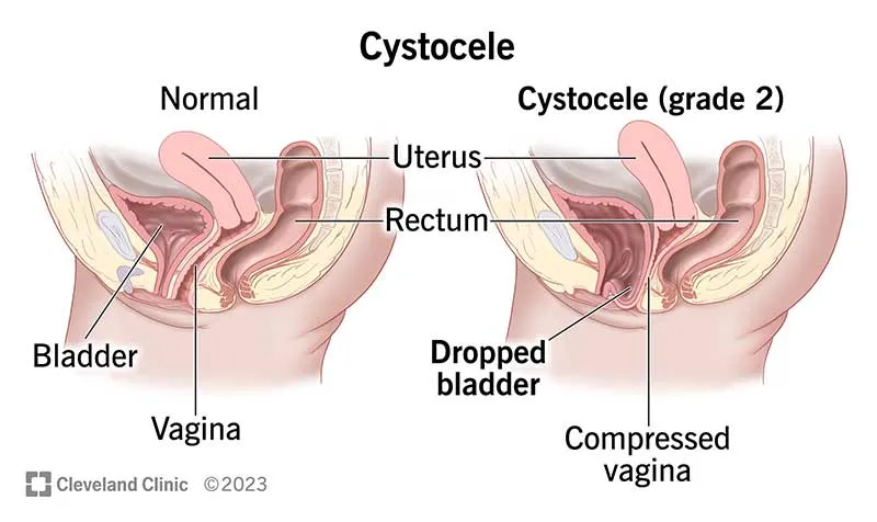
The vagina and uterus are supported by a network of supportive fasciae (such as paravaginal fascia) and pelvic floor muscles. When these fasciae become weak or torn, or when the pelvic floor muscles are paralyzed or stretched, the intra-abdominal pressure is no longer evenly distributed. Over time, this causes the walls to become lax and protrude. Types of prolapse, based on the dominant location, include anterior prolapse (cystocele), apical prolapse (vaginal or uterine vault prolapse), and posterior prolapse (rectocele), which are often seen together.

عمل جراحي ركتوپكسي براي پرولاپس ركتوم

Manifestations

  • Feeling of Pressure, Heaviness, or a “Something” Coming Out of the Vagina: Sometimes the patient can see or feel this bulge.
  • Feeling of Incomplete Evacuation or the Need for Manual Pressure for Rectal Emptying (if rectocele is present).
  • Urinary Problems: Frequency, urgency, or stress incontinence (urine leakage during coughing/laughing) or urinary retention in apical prolapse.
  • Pain or Discomfort During Sexual Intercourse (dyspareunia).
  • Occasional Low Back Pain or Discomfort in the Perineal Area.

Diagnosis

A) Detailed Medical History

Questions the doctor may ask: Duration and intensity of pain, bowel movement sequence, constipation or diarrhea, history of pregnancy/childbirth, use of anticoagulant medications, warning signs (weight loss, fever, heavy bleeding, systemic symptoms). This information is crucial for determining the need for further investigations.

B) Clinical and Visual Examination

A thorough examination of the skin around the anus typically provides the diagnosis; fissures are most commonly seen along the posterior midline. Digital rectal examination (DRE) is only performed if the patient can tolerate it or under anesthesia.

C) Anoscopy / Proctoscopy

After initial pain relief, simple anoscopy helps visualize the position of the fissure, associated papilla, or hemorrhoids. Proctoscopy is used to examine higher areas.

D) When is Colonoscopy Necessary?

If the fissure is: non-midline, multifocal, resistant to treatment, or accompanied by warning signs (weight loss, long-term changes in bowel habits) → colonoscopy is essential to rule out inflammatory bowel disease or higher lesions. This is important because not all fissures require colonoscopy, but in unusual cases or for patients over 40, a full colon evaluation is recommended.

E) Anorectal Manometry (in Specific Cases)

In patients who are to undergo treatments that stimulate tone (such as Botox) or who need an evaluation of incontinence before surgery, anorectal manometry will assess resting pressure and sphincter contraction, aiding in decision-making.

Treatment of Uterine and Vaginal Prolapse

A) Conservative Treatment

  1. Pelvic Floor Muscle Training (PFMT)

  • Scheduled and guided by a physiotherapist; correct technique and follow-up are crucial. Results are generally better than self-directed exercises.
  1. Pessary

  • A silicone or plastic device placed inside the vagina to provide mechanical support and reduce symptoms.
  • Suitable for women who are not willing to undergo surgery, older women, or those who wish to try non-surgical options before making a final decision.
  • Care: Fitting, training on replacement, or regular replacement visits are necessary. If pain or discharge occurs, medical consultation is required.
  1. Topical Estrogen for Postmenopausal Women

  • Improves the quality of mucosal and vaginal tissue, alleviating symptoms related to atrophy.

These methods can be particularly beneficial in mild to moderate cases and for maintaining quality of life.

B) Surgical Treatment

  1. Vaginal Repair (Approaches via Vagina)

  • Anterior/Posterior Repair: For cystocele or rectocele, performed directly via the vagina. Advantages: No abdominal incision, shorter recovery time. Limitations: In some cases, shorter durability and higher recurrence rates compared to abdominal approaches in the long term.
  • Manchester Procedure
  1. Abdominal/Laparoscopic Surgeries — Sacrocolpopexy / Sacrohysteropexy

  • Performed via laparoscopy or robotic surgery, where the vaginal or uterine vault is fixed to the sacrum with mesh to provide long-term support.
  • Advantages: Higher durability, lower recurrence rates, better anatomical visualization, and more precise tissue support. Laparoscopic or robotic methods offer less pain, quicker recovery, and potentially better pelvic nerve protection. Due to these benefits, this approach is often preferred in specialized centers.
  • Note on Mesh: Mesh use should be approached with caution and based on the experience of the center. The benefits and risks must be clearly communicated with the patient.
  1. Uterine-Sparing vs. Hysterectomy

  • For patients who wish to preserve their uterus or need to do so for medical reasons, options such as sacrohysteropexy are available, providing abdominal support for the uterus.
  • In other cases, a hysterectomy (removal of the uterus) may be performed along with vaginal vault fixation. The choice depends on the patient’s preference, uterine pathology (e.g., fibroids, bleeding), and anatomical factors.
  1. Combination and Multi-Organ Corrections

  • Many patients present with multi-compartment prolapse (e.g., cystocele + rectocele + apical prolapse). Effective correction usually requires planning to address all areas of weakness to achieve a stable result.

Role of colorectal Surgeon

  • Clear and Comprehensive Explanation of Options, Benefits, and Risks: The colorectal surgeon provides detailed information about the available treatment options, their advantages, and potential risks, helping the patient make an informed decision.
  • Choosing the Appropriate Method: The surgeon selects the best treatment approach based on the patient’s age, desire to preserve the uterus or plan for future pregnancies, and imaging findings.
  • Collaboration Among Specialties: The colorectal surgeon plays a key role in coordinating with other specialists, including pelvic surgeons, urogynecologists, pelvic floor physiotherapists, and urologists to ensure a comprehensive treatment plan.
  • In Specialized Centers: In well-equipped centers, the colorectal surgeon uses laparoscopic or robotic surgery and neuroprotective techniques to minimize the risk of urinary or sexual dysfunction.

Frequently Asked Questions about Uterine and Vaginal Prolapse

Do I Always Need Surgery?

No. If the symptoms are mild, conservative options like pessary use and pelvic floor exercises are suitable. Surgery is recommended when symptoms affect daily life or sexual relations, or if non-surgical treatments are not effective.

A pessary is a silicone or plastic device that is inserted into the vagina to provide support. The doctor will measure the appropriate size and provide instructions for replacement or regular follow-up visits for care.

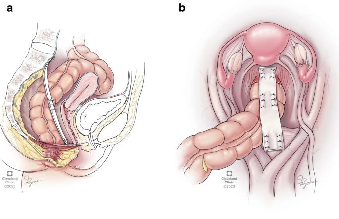
Laparoscopy or robotic surgery offers similar oncological outcomes, less pain, faster recovery, and better nerve protection. These techniques are especially recommended for abdominal repairs or sacrocolpopexy in specialized centers.

Initial Post-Operative Care Relative Rest: Take it easy initially, avoiding prolonged straining and heavy lifting for a few weeks. Pelvic Floor Physiotherapy: Recommended if advised by your doctor to aid recovery. Follow-Up Appointments: Attend scheduled follow-up visits as instructed by your healthcare provider to monitor your recovery.

Summary for Patient

Uterine and vaginal prolapse refers to the descent of pelvic structures, which can cause feelings of pressure, urinary problems, and discomfort during intercourse. Treatment options vary from exercises and pessary use to vaginal or abdominal surgeries (laparoscopic/robotic). The best treatment approach is chosen based on the severity of symptoms, your preference for preserving the uterus, and examination and imaging findings. In specialized centers, minimally invasive methods performed by experienced surgeons typically yield more durable results.