Cecal Cancer

سرطان های روده بزرگ

Introduction

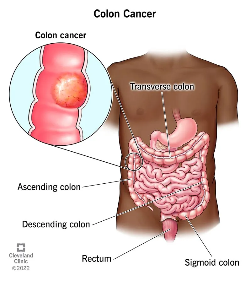
Cecal cancer refers to the development of a malignant (cancerous) tumor in the cecum, which is the first portion of the large intestine located in the lower right quadrant of the abdomen—where the small intestine (ileum) connects to the large intestine. In simple terms, it is a tumor that arises from the cells of the cecal wall and may extend into deeper layers of the bowel, spread to surrounding tissues, or disseminate to distant sites through the bloodstream or lymphatic system.

Epidemiology (Prevalence and Risk Factors)

Cecal cancer, as part of right-sided colon cancers, accounts for approximately 10–15% of all colorectal malignancies. It occurs more frequently in older individuals and is often diagnosed through findings such as iron-deficiency anemia or occult gastrointestinal bleeding. According to global statistics, colorectal cancer is the second most common cancer in women and the third in men, with more than 1.9 million new cases reported annually worldwide. In Iran, colorectal cancer represents about 9–10% of all cancers and is considered one of the five most common malignancies, with an increasing trend in recent years. The exact proportion of cecal cancer within national data is not precisely defined, but most studies indicate a distribution similar to global patterns.

سرطان روده بزرگ

Etiology (Causes and Disease Mechanisms)

  • Genetic factors and family history: Having a parent or sibling with the disease may increase the risk.
  • Adenomatous polyps (precancerous polyps): If not removed, they can gradually transform into cancer over the years.
  • Advanced age and lifestyle factors: High-fat, low-fiber diets, obesity, and physical inactivity contribute to increased risk.
  • Inflammatory bowel disease: Long-standing conditions such as ulcerative colitis raise the likelihood of developing cancer.
  • Dietary habits and alcohol/tobacco use: Chronic alcohol consumption, smoking, and an unhealthy diet have been associated with elevated risk.

Pathogenesis (Mechanisms of Disease Development)

  • The process typically begins with an adenomatous polyp—a small benign growth protruding from the mucosal surface. Over several years, some of these polyps acquire genetic mutations and transform into malignant cells.
  • As the cancer progresses, malignant cells may invade deeper layers of the bowel wall, reach lymphatic or blood vessels, and spread to nearby lymph nodes or distant organs such as the liver or lungs.
  • Some tumors exhibit specific molecular characteristics that influence treatment decisions, including their responsiveness to targeted therapies or immunotherapy.

Clinical Manifestations

Cecal cancer may remain asymptomatic for a long time because the cecum has a relatively large diameter, allowing the tumor to grow without causing obstruction. This means symptoms may be mild or subtle:

برداشتن روده
  • Fatigue and weakness due to anemia (often iron-deficiency anemia) — caused by subtle, chronic bleeding from the tumor. The patient may not notice visible blood in the stool and instead experiences only low iron levels and fatigue.
  • Loss of appetite or unintentional weight loss.
  • Dull or vague discomfort in the lower right abdomen — which may be mild in early stages.
  • Changes in bowel habits, such as diarrhea, constipation, or a sensation of incomplete evacuation.
  • Overt bleeding (occasionally) or blood in the stool — although in cecal cancer, bleeding may present as dark stools or may be clinically occult and not visible to the patient.

In advanced cases: a palpable mass in the lower right abdomen, signs of bowel obstruction (nausea, vomiting, abdominal distension), or symptoms related to metastases—such as hepatic pain or shortness of breath if the liver or lungs are involved.

Important note: New-onset iron-deficiency anemia in an adult without an identifiable cause should prompt evaluation for a gastrointestinal source of bleeding, including colorectal cancer.

Diagnosis

1.Colonoscopy with Biopsy

The most important and definitive diagnostic method. Using a flexible, camera-equipped scope, the physician visualizes the colon and obtains samples from any suspicious lesion. Pathology (microscopic examination) determines whether the cells are malignant.

2.Blood Test — CEA (Carcinoembryonic Antigen)

CEA is a tumor marker that may be elevated in some patients. An increased level is not diagnostic on its own but is useful alongside other clinical information and for post-treatment monitoring.

3.Imaging for Staging

Contrast-enhanced CT of the abdomen and pelvis is used to assess tumor extent, lymph node involvement, and possible liver metastases; chest CT is often performed to evaluate the lungs. In selected cases, MRI or PET-CT may also be used. These imaging studies determine whether the disease is localized or has spread, and are essential for treatment planning.

4.Molecular Testing (when indicated)

Assessment of the tumor’s genetic features (such as MSI status or specific gene mutations) can help guide the choice of chemotherapy, targeted therapies, or immunotherapy. In simple terms: tests that indicate which treatments are likely to be more effective.

Treatment of Cecal Cancer

Overall goal: to remove the tumor, prevent further spread, and improve survival and quality of life.

A — Surgery

  • The standard operation for cecal cancer is typically a right hemicolectomy.
  • In simple terms, the surgeon removes the cecum, the initial portion of the ascending colon, and sometimes part of the ileum (the terminal segment of the small intestine), then connects the healthy ends of the bowel together (an anastomosis).
  • Along with removing the involved bowel segment, the surgeon also removes the adjacent lymph nodes for pathological evaluation and to reduce the risk of local spread. Adequate lymph node retrieval (usually at least 12 nodes) is recommended to ensure accurate staging.
  • Surgical approach: The procedure may be performed as an open surgery (large incision) or via minimally invasive techniques (laparoscopic or robotic). When available, laparoscopic surgery offers advantages such as smaller incisions, less postoperative pain, and faster recovery. The choice of technique depends on the patient’s condition and the experience of the surgical center.
  • Timing of surgery: If the patient presents emergently with obstruction or perforation, urgent intervention may be necessary, and staged procedures (such as temporary stoma creation) may sometimes be required. In elective (planned) settings, surgery is performed after appropriate preoperative preparation.
برداشتن روده

B — Chemotherapy

  • In some tumors with specific molecular characteristics, targeted therapies or immunotherapy may be added or used as alternatives. This decision is made based on the tumor’s molecular profile and the recommendations of the oncology team.

C — Supportive and Palliative Care

  • In advanced stages, the focus is often on symptom control, improving quality of life, nutrition, and pain relief.

Role of the Colorectal Surgeon

  • Complete tumor removal with clear margins: The surgeon must excise the tumor along with a margin of healthy surrounding tissue to minimize the risk of local recurrence.
  • Adequate lymph node dissection: The number and status of the lymph nodes removed are essential for accurate staging and for planning postoperative treatment.
  • Preservation of bowel function: When feasible, the surgeon aims to restore bowel continuity directly so the patient does not require a stoma; however, in complex or emergency situations, creation of a stoma may be necessary.
  • Coordinated multimodal treatment planning: The surgeon is part of a multidisciplinary cancer team—working with oncologists, pathologists, radiologists, and specialized nurses to determine the need for chemotherapy or other treatments.
  • Clear communication with the patient: The surgeon must explain the goals of surgery, potential risks, and possible outcomes (such as changes in bowel habits, postoperative pain, infection risk, or recurrence).

Frequently Asked Questions About Cecal Cancer

Is cecal cancer treatable?

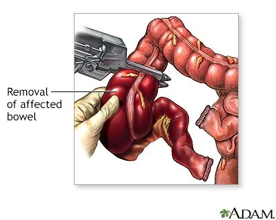
Yes — if detected early and the tumor is confined to the cecum, surgery with or without chemotherapy offers a high chance of cure. However, each case must be evaluated individually.

If a first-degree relative (parent, sibling) has been affected, or if you have a history of multiple polyps or a genetic condition, genetic counseling and a specialized screening plan are recommended. Otherwise, the general recommendation is for every adult to undergo routine colorectal screening—such as colonoscopy or other approved screening tests—according to local guidelines.

No; many gastrointestinal conditions can cause these symptoms, but any new or concerning symptom should be medically evaluated to determine the exact cause.

Right hemicolectomy generally does not have a significant impact on fertility in women (unlike surgeries performed deep within the pelvis). However, if you have concerns, you should discuss them with the surgical team before the operation so that appropriate planning can be made.

Depending on the surgical approach (laparoscopic or open), your overall condition, and the presence of any complications, recovery usually takes from several weeks to a few months. The surgical team will guide you regarding the appropriate time to return to work and resume normal daily activities.

Summary for Patient

Cecal cancer is a tumor that develops in the first part of the large intestine. It may begin with fatigue and anemia, and in more advanced stages can present with pain or changes in bowel habits. Definitive diagnosis is made through colonoscopy and biopsy; surgery is the main treatment, with chemotherapy or targeted therapies added in certain stages. The earlier the tumor is detected, the better the chance of recovery. If you experience unexplained fatigue, weight loss, or gastrointestinal bleeding, be sure to consult a physician.