Functional Constipation

Introduction

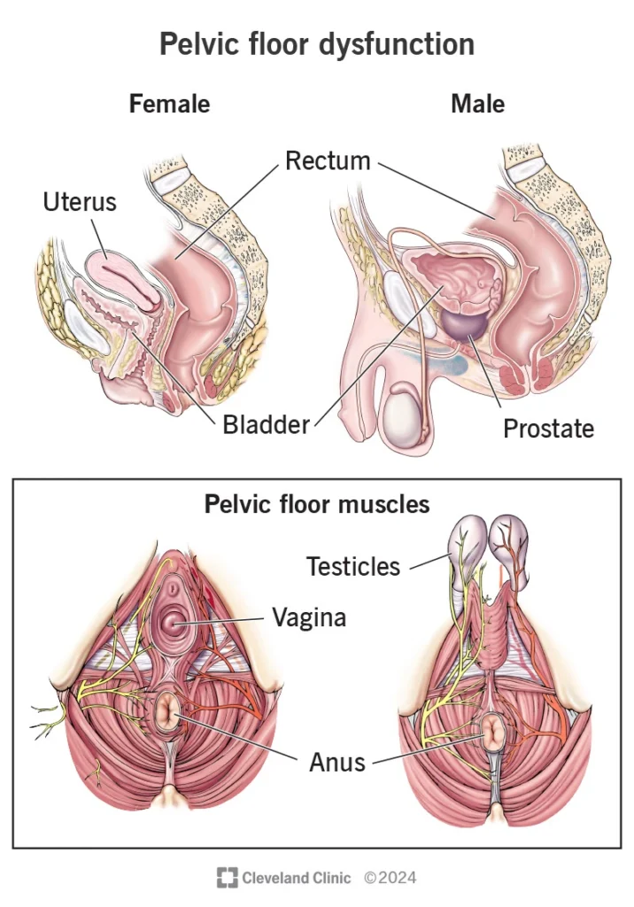
Functional constipation, also known as obstructed defecation syndrome (ODS), refers to a condition in which a patient is unable to effectively empty the rectum despite feeling the urge to defecate.
The hallmark symptoms include prolonged straining, a persistent sense of incomplete evacuation, the need for manual assistance (digitation) to help pass stool, and spending excessive time on the toilet.

In this syndrome, both functional problems of the pelvic floor muscles (such as dyssynergia) and anatomical abnormalities—including rectocele, mucosal prolapse, or intussusception—may be present.
Therefore, diagnosis and treatment must be multidisciplinary and combined, addressing both functional and structural components.

Epidemiology (Prevalence and Risk Factors)

ODS is one of the common forms of chronic constipation.
Although prevalence varies across studies in developed countries, a significant proportion of patients with chronic constipation—up to 20–30% in some populations—report symptoms of inadequate evacuation or excessive straining. The condition is more frequent in women, particularly those with a history of childbirth, and is seen more often in middle-aged and older adults.

Etiology (Causes and Pathophysiology)

1. Functional (Non-Anatomical) Factors

  • Dyssynergia, or lack of coordinated relaxation of the pelvic floor muscles and anal sphincter during defecation (anismus)

  • Weakness or poor coordination of the pelvic floor muscles, and abnormal rectal sensation

2. Anatomical Factors

  • Rectocele (bulging of the anterior rectal wall toward the vagina), which can cause stool trapping.
  • Internal intussusception or mucosal prolapse, where part of the rectal wall folds inward and obstructs stool passage.
  • Perineal descent or pelvic floor laxity, and partial or full-thickness rectal prolapse.
  • Changes related to difficult childbirth (such as sphincter tears or damage to supporting tissues), obesity, a history of pelvic surgery, or nerve injury.

In many patients, a combination of multiple factors occurs simultaneously, which is why a comprehensive evaluation is essential.

Pathogenesis (Mechanism of Disease Development)

  • In dyssynergia, the pelvic floor muscles and anal sphincters fail to relax or coordinate properly during attempts to defecate. Instead of opening, they may paradoxically contract, blocking stool passage.
  • In rectocele, the anterior rectal wall bulges toward the vagina, creating a pocket where stool becomes trapped, making evacuation difficult.
  • In internal intussusception or mucosal prolapse, the rectal wall “folds into itself,” narrowing or obstructing the outlet and preventing effective stool passage.

All of these mechanisms may cause symptoms individually or in combination, which is why a simultaneous assessment of both functional and anatomical factors is essential.

Clinical Presentation

  • Prolonged straining with ineffective evacuation
  • A persistent sensation of incomplete emptying or stool remaining in the rectum
  • The need for manual assistance (applying pressure on the perineum or using a finger in the vagina or anus)
  • The need for excessive force or spending more than 10–15 minutes to defecate
  • Repeated toilet attempts, bloating, or even mucus leakage
  • In mixed cases, mild fecal or gas incontinence may also be present

Diagnosis

Diagnosis of ODS requires a combination of history-taking, physical examination, and functional/imaging studies.
The usual sequence and practical considerations are as follows:

1) Structured History and Questionnaires

  • Questionnaires such as the ODS score and bowel diaries help quantify the severity of symptoms and monitor treatment progress.

2) Physical Examination and Anal/Vaginal Examination

  • Assessment includes sphincter tone, identification of rectocele or visible prolapse during Valsalva straining, and evaluation for obstetric trauma (such as scarring or sphincter defects).

3) Functional Tests (Based on Clinical Findings)

  • Balloon Expulsion Test: A small balloon is placed in the rectum, and the patient attempts to expel it while seated as if on a toilet. The time required reflects rectal evacuation ability. No preparation is needed, and the test is performed in the clinic.
  • Anorectal Manometry:Measures resting and squeeze pressures of the sphincters and evaluates pelvic floor coordination. This test helps diagnose dyssynergia. It typically takes 20–30 minutes and requires no special preparation.
  • Endoanal Ultrasound (EAUS): Evaluates sphincter structure (tears or scarring), especially in cases of childbirth-related or postsurgical injury. Performed in the clinic and essential for planning sphincter repair.

4) Dynamic Imaging — To Visualize Actual Defecation Mechanics

  • Fluoroscopic Defecography: After the rectum is filled with a stool-like contrast paste, the patient sits on a specialized commode while X-ray fluoroscopy captures defecation in real time. This shows rectocele size, internal intussusception, perineal descent, and anorectal angle motion. Preparation typically includes an enema or rectal filling as instructed by the center.
  • MRI Defecography: Similar to fluoroscopic defecography but with superior soft-tissue detail, simultaneous visualization of all three pelvic compartments (e.g., enterocele), and no ionizing radiation. Although studies show no universal superiority of one method over the other, MRI offers clearer soft-tissue imaging and greater patient comfort. Preparation involves rectal filling with appropriate material and detailed explanation of the maneuvers.

Treatment of Functional Constipation (ODS)

(Step-by-Step Management Guide)

Before any invasive intervention—especially surgery—the patient should undergo at least three months of medical therapy and defecation retraining, including biofeedback.

1) Basic Treatments and Lifestyle Modification

  • Gradual fiber intake up to 20–30 grams per day (rapid increases may cause bloating; slow titration is preferred).
  • Adequate hydration.
  • Stool softeners (such as polyethylene glycol) or osmotic laxatives under medical supervision when stool is hard.
  • Optimized toileting behavior: proper timing, correct sitting posture, breathing techniques, and coordinated abdominal pressure rather than sudden straining.

2) Pelvic Floor Rehabilitation and Biofeedback (The First Specialized Step)

  • The goal of biofeedback is to restore proper coordination of the pelvic floor muscles.
    Using pressure sensors or EMG, the patient sits in front of a monitor and learns when to relax the pelvic floor and how to generate appropriate abdominal pressure during defecation.
  • A typical program includes 6–12 sessions, held weekly or biweekly over several weeks, along with home exercises.
    Randomized trials and meta-analyses show that biofeedback leads to significant improvement in symptoms and functional outcomes in patients with dyssynergia, with demonstrated long-term efficacy.

3) Medical and Adjunctive Therapies

  • Laxatives, stool softeners, and medications that regulate bowel motility may be used as needed.
  • Antispasmodic or analgesic medications can be prescribed in selected cases.
  • Neuromodulation techniques—such as tibial nerve stimulation or sacral neuromodulation—have been investigated in some centers and may be helpful for selected patients, either within research settings or after failure of other treatments (evidence varies).

4) Surgical Options

A) STARR (Stapled Transanal Rectal Resection)
  • Through the anus, a stapling device is used to remove redundant mucosal and submucosal tissue responsible for bulging, rectocele, or internal mucosal prolapse.The area is then reconstructed using sutures and staples.
  • Indications: A large rectocele or internal mucosal prolapse that represents the primary mechanical issue, in a patient with adequate sphincter function.
  • Advantages / Limitations: Provides early symptom relief; short-term improvement is reported in 70–90% of patients. However, long-term outcomes show recurrence rates of 30–40%, emphasizing the need for careful patient selection. Rare but significant complications—such as major bleeding or persistent pain—have been reported.

B) Laparoscopic / Robotic Ventral Mesh Rectopexy (LVMR / RVMR)
  • Using a laparoscopic or robotic approach from the abdomen, the anterior rectum is mobilized and secured with a lightweight mesh to the posterior wall of the uterus or bladder, thereby stabilizing the upper rectum.
    During the same procedure, a rectocele or internal intussusception can also be corrected.
  • Indications: Full-thickness rectal prolapse, large rectocele, or internal intussusception that requires anatomical correction to restore proper pelvic support and function.
C) Perineal and Transvaginal Approaches (For Selected Patients)
  • Procedures such as transvaginal rectocele repair or the Altemeier/Delorme techniques for specific types of rectal prolapse may be used in carefully selected patients.
    The choice of approach depends on the patient’s overall condition, anatomical characteristics, and surgical risk.
D) Choosing the Treatment Method Based on the Primary Cause
  • If the main problem is functional (such as dyssynergia) → the focus should be on biofeedback and pelvic floor physiotherapy, and surgery should be avoided initially.
  • If there is a significant and correctable anatomical defect (such as a large rectocele or full-thickness prolapse) with clear corresponding symptoms → anatomical surgical repair (such as LVMR or other appropriate techniques) is indicated.

E ) Alternative Surgical Approaches

(For patients at risk of incontinence or when LIS is not appropriate)

  • Mucosal or anocutaneous advancement flap:
    This technique involves excising the fibrotic tissue and covering the defect with healthy flap tissue.
    It is particularly suitable for patients with low sphincter tone or those at high risk of postoperative incontinence.
  • Customized or tailor-made combinations of techniques may also be used, depending on clinical examination findings and the patient’s history of childbirth or sphincter injury.

Role of the Colorectal Surgeon

  • Technical decision-maker: reviewing MRI/defecography and manometry results, and determining whether surgery is indicated and which surgical approach is most appropriate.
  • Performer of specialized procedures such as LVMR, STARR, perineal operations, and sphincter repair when needed, as well as managing postoperative complications.
  • Team collaboration: working closely with pelvic floor physiotherapists, radiologists, and—when treating women with associated prolapse—sometimes with urologists or gynecologists.
  • Patient education and follow-up: guiding patients before and after surgery, managing expectations, and coordinating rehabilitation.

Frequently Asked Questions About Functional Constipation (ODS)

1) Do all rectoceles require surgery?

No. Small rectoceles without symptoms—or those causing only mild constipation—are usually managed with medical therapy and pelvic floor physiotherapy.
Surgery is considered only when the rectocele is large, clearly associated with incomplete evacuation, and unresponsive to biofeedback.

Biofeedback typically involves 6–12 sessions, held weekly or every two weeks, along with home exercises.
Many patients notice early improvement after just a few sessions, but completing the full course is important for long-term skill reinforcement.
Evidence shows that biofeedback is effective and is considered the first-line treatment for dyssynergia.

Both have advantages and limitations.
Fluoroscopic defecography is less expensive, provides real-time imaging, and is very useful for assessing evacuation mechanics.
MRI defecography avoids radiation, offers superior soft-tissue detail, and visualizes all three pelvic compartments simultaneously.

Studies show no absolute superiority of one method over the other; the choice depends on availability and the experience of the center performing the test.

The next step depends on imaging and manometry findings.
If a correctable anatomical problem is present, a targeted surgical procedure—such as LVMR or STARR in selected cases—may be considered.
These decisions are made within a multidisciplinary team to ensure the most appropriate and effective treatment plan.

The goal of surgery is to improve quality of life and make evacuation easier.
Some patients experience significant improvement, while others achieve only partial relief.
Proper patient selection and the experience of the surgeon play key roles in determining the final outcome.

Summary for Patients

Functional constipation, or ODS, refers to difficulty emptying the bowels, which may result from pelvic floor muscle dysfunction or from anatomical problems such as rectocele or prolapse.
The first and most important step in management is always non-surgical therapy and rehabilitation, particularly biofeedback.Dynamic imaging and functional tests help identify the primary cause. Surgical options are considered only when conservative treatments fail and a correctable anatomical abnormality is present. Decision-making should involve a multidisciplinary team—including an experienced colorectal surgeon, a radiologist, and a pelvic floor physiotherapist—to ensure the best possible outcome.